Pennisetum glaucum (Pearl Millet) Extract Exhibits Anticoagulant, Antiplatelet, Antioxidant and Proteolytic Activity
Devaraja Sannaningaiah1*, Ashwini Shivaiah1, Jayanna Kengaiah1, Sharath Kumar M Nandish1, Chethana Ramachandraiah1, Chandramma1, Sebastin Santosh M1, and Thippeswamy Thippande Gowda1 and Manohar Shinde2
1Department of Biochemistry, Tumkur University, Tumkur, India
2Department of Medicinal Biochemistry and Microbiology, Uppsala biomedical Centre, Uppsala, Sweden
- *Corresponding Author:
- Devaraja Sannaningaiah
Department of Studies and Research in Biochemistry,
Tumkur University,
Tumkur,
India;
Email: sdevbiochem@gmail.com
Received: December 23, 2019, Manuscript No. IPJCND-23-3109; Editor assigned: December 26, 2019, PreQC No. IPJCND-23-3109 (PQ); Reviewed: January 10, 2019, QC No. IPJCND-23-3109; Revised: October 03, 2023, Manuscript No. IPJCND-23-3109 (R); Published: October 31, 2023, DOI: 10.36648/2472-1921.9.10.81
Citation: Sannaningaiah D, Shivaiah A, Kengaiah J, Nandish SKM, Ramachandraiah C, et al. (2023) Pennisetum glaucum (Pearl Millet) Extract Exhibits Anticoagulant, Antiplatelet, Antioxidant and Proteolytic Activity. J Clin Nutr Die Vol.9 No.10: 81.
Abstract
Background: Thrombotic Disorders (TD) increase the risk of cardiovascular/cerebrovascular complications and represent a major health problem worldwide. Although, anticoagulant, antiplatelet and fibrinolytic agents have been widely used, their life threatening side effects limits their usage.
Objective: The objective of this study is to evaluate the protective role of Pennisetum glaucum Extract (PGE) on thrombotic disorder.
Methods: Both in-vitro and in-vivo experiments were carried out to establish the anticoagulant, antiplatelet and antioxidant properties of PGE. The parameters such as plasma recalcification time, Prothrombin Time (PT), Activated Partial Thromboplastin Time (APTT) and agonist induced platelet aggregation assays were employed. PGE was also analysed for its efficacy on oxidative stress as well.
Results: PGE exhibited strong anticoagulant property in both Platelet Rich Plasma (PRP) and Poor Plasma (PPP). PGE was also exhibited anticoagulant effect only in Activated Partial Thromboplastin Time (APTT) but not in Prothrombin (PT), revealed its interference in intrinsic pathway of blood coagulation cascade. PGE displayed antiplatelet property by inhibiting platelet aggregation process induced by agonists such as ADP and epinephrine. PGE significantly normalized the level of lipid peroxidation, protein carbonyl content and total thiol in the sodium nitrate treated hemolysates. PGE was devoid of haemolytic, edematic and hemorrhagic activities.
Conclussion: PGE showed zinc dependent proteolytic activity, anticoagulant, antiplatelet, antioxidant and nontoxic properties. Hence, it could be a valuable candidate in the management of thrombotic complications.
Keywords
Pearl millet; Anticoagulant; Antiplatelet; Protease; Hemolysates
Introduction
An increasing life-expectancy and world population explosion has been associated with communicable and non-communicable diseases worldwide. While, oxidative stress is the key contributing factor involved in the pathogenesis of atherosclerosis, hypertension, diabetes mellitus, ischemic diseases, malignancies, and thrombosis. Cardiovascular disease is a major non-communicable disease leading to death and disability [1]. The key pathology involved in three major cardiovascular disorders such as, ischemic heart disease, stroke and venous thromboembolism is the thrombosis. Thrombosis is nothing but the formation of unusual clot in veins and arteries, involves unregulated way of activation of several coagulation factors and platelets. Target specific anticoagulant and antiplatelet therapy is the currently available remedy [2]. Several synthetic and natural drugs are available in the market; however, their life threatening side effects (internal bleeding, nausea, miscarriage and vomiting) limits their usage. Therefore, identification of novel anticoagulant and antiplatelet drug with zero side effects from the natural sources is the big challenge to the researchers. Since ancient times, natural products from animals, microbes, plants and marine source have been extensively used in the treatment of several diseases. The wisdom of our ancestors on folk medicine is the base for modern drug discovery process [3].
The key thirst for scientist to look into natural product could be due their tremendous chemical and structural diversity. According to the documentation by WHO about 80% of the world’s population is devoted to folk medicine prepared from the plants for primary health care. Recent data suggests that only 5%-15% of terrestrial plant species have been explored pharmacologically. About 25% medicines which are commercially available today in the market are derived from plants origin. Morphine was the first commercialized plantderived medicine isolated from Papaver somniferum. Subsequently several compounds have been isolated and commercialized [4]. As life style diseases tremendously increasing especially in developing countries, there has been a food revolution over the past few years. Thus, people are rethinking about using millets as the major staple food by replacing ragi, rice and wheat. Though, Pennisetum glaucum (pearl millet) has been widely cultivated in Africa and Asian continent, India is the largest producer. Pennisetum glaucum are cereal crops they have been widely used from prehistoric period due to their umpteen health benefits. They are the richest source of macro and micro nutrients with high quantity of dietary fibers. They found to exhibit immense health benefits as they are capable of curing coronary heart diseases, diabetes, constipation, jaundice and high blood pleasure. Despite, immense therapeutic applications of Pennisetum glaucum, its beneficial role on thrombosis and oxidative stress was not explored. Thus, the current study aims to identify the beneficial role of protein constituents of Pennisetum glaucum (pearl millet) on thrombosis and oxidative stress [5,6].
Materials and Methods
Fatâ??free casein, gelatine, Phenyl Methyl Sulphonyl Fluoride (PMSF), Ethylene Di-amine Tetra-acetic Acid (EDTA), Iodo-Acetic Acid (IAA), and 1,10â??Phenanthroline were purchased from Sigma chemicals company (St. Louis, USA). Molecular weight markers were from Bangalore Genie Private Limited, India. Activated Partial Thromboplastin Time (APTT) and Prothrombin Time (PT) reagents were purchased from AGAPPE Diagnostic Pvt., Ernakulum, Kerala, India. Human plasma fibrinogen was purchased from Sigma chemicals Co., St. Louis, USA. All other chemicals used were of analytical grade [7]. Fresh human blood was collected from healthy donors for the Plateletâ??Rich Plasma (PRP) and Platelet poor Plasma (PPP). ADP and Epinephrine were purchased from Sigma chemicals company (St. Louis, USA). All the experimentations were conducted in accordance with the ethical guidelines and were approved by the Institutional Human Ethical Committee (IHEC-UOM No. 47Res/2014-15), University of Mysore, Mysore. Conducting animal experiments were permitted by the Institutional Animal Ethical Committee (UOM/ IAEC/02/2016), University of Mysore [8]. The animal handling were proceeded in accordance with the guidelines of the Committee for the Purpose of Monitoring and Supervision of Experiments on Animals (CPCSEA).
Preparation of Pennisetum Glaucum Extract (PGE) and Protein estimation
Pennisetum glaucum was purchased from local market Tumkur. The dried seeds were powdered, dissolved in double distilled water and centrifuged at 8000 g for 15 min, collected the supernatant and stored in freezer at -20°C until further usage. Protein concentration was determined as described by Bradford et al. Using Bovine Serum Albumin (BSA) as standard [9,10].
Sodium Dodecyl Sulphate Polyacrylamide Gel Electrophoresis (SDS-PAGE) and Periodic Acid-Schiff (PAS) staining
Sodium Dodecyl Sulphate-Polyacrylamide Gel Electrophoresis (SDS-PAGE) 10% was carried out according to the method of Laemmli. The crude PGE (100 μg) and PGE (50 μg) prepared under reducing and non-reducing conditions was used for SDSPAGE. The electrophoresis was carried out using Tris (25 mM), glycine (192 mM) and SDS (0.1%) for 2 hr at room temperature. After electrophoresis, the SDS-PAGE gels were stained with 0.1% Coomassie brilliant blue R-250 for detection of the protein bands and de-stained with 40% ethanol in 10% acetic acid and water (40:10:50v/v). Molecular weight standards from 200 kDa to 14. 3kDa were used [11].
PAS staining
PAS staining was carried out according to the method of Leach, et al. after electrophoresis; the gel was fixed in 7.5% acetic acid solution and stored at room temperature for 1 hr. Then the gel was washed with 1% nitric acid solution and kept in 0.2% aqueous periodic acid solution and stored at 4°C for 45 min. After that, the gel was placed in Schiff’s reagent at 4°C for 24 hr and was de-stained using 10% acetic acid to visualize a pink colour band [12].
Proteolytic activity
Proteolytic activity was assayed as described by Satake, et al. fat-free casein (0.4 ml, 2% in 0.2 M Tris-HCl buffer, pH 7.6) was incubated with 10 μg of PGE in a total volume of 1 ml for 2 hr and 30 min at 37°C. Undigested casein was precipitated by adding 1.5 ml of 0.44 M Trichloroaceticacid (TCA) and left to stand for 30 min. The reaction mixture was then centrifuged at 2000 g for 10 min. Sodium carbonate (2.5 ml, 0.4 M) and Folin- Ciocalteu’s reagent (1:2) were added sequentially to 1 ml of the supernatant and the colour developed was read at 660 nm. One unit of the enzyme activity was defined as the amount of the enzyme required to cause an increase in Optical Density (OD) of 0.01 at 660 nm. The specific activity was expressed as units/min/mg of protein. For inhibition studies, a similar reaction was performed independently after pre-incubating the PGE (10 μg) for 30 min with 5 mM each of EDTA, 1,10- phenanthroline, PMSF and IAA. In all the cases, appropriate controls were kept [13].
Plasma re-calcification time
The plasma re-calcification time was determined according to the method of Quick, et al. Briefly, the PGE (2 μg-14 μg) was preincubated with 0.2 ml of citrated human plasma in the presence of 10 mM Tris HCl (20 μl) buffer pH 7.4 for 1min at 37°C, 20 μl of 0.25 M CaCl 2 was added to the pre-incubated mixture and clotting time was recorded.
APTT and PT
Briefly, 100 μl of normal citrated human plasma and PGE (0-10 μg) were pre-incubated for 1 min. For APTT, 100 μl reagent (LIQUICELIN-E Phospholipids preparation derived from rabbit brain with ellagic acid), which was activated for 3 min at 37°C was added. The clotting was initiated by adding 100 μl of 0.02 M CaCl 2 and the clotting time was measured. For PT, the clotting was initiated by adding 200 μl of PT reagent (UNIPLASTIN-rabbit brain Thromboplastin). The time taken for the visible clot was recorded in seconds. The APTT ratio and the International Normalized Ratio (INR) for PT at each point was calculated from the values of control plasma incubated with the buffer for identical period of time.
Fibrinogenolytic activity
Fibrinogenolytic activity was determined as described previously by Ouyang and Teng et al. PGE (0 μg-10 μg) was incubated with the human plasma ibrinogen (50 μg) in a total volume of 40 μl of 10 mM Tris-HCl buffer pH 7.4 for 4 hr at 37°C. A ter the incubation period, reaction was terminated by adding 20 μl denaturing buffer containing 1 M urea, 4% SDS and 4% β- mercaptoethanol. It was then analysed by 10% SDS-PAGE.
Degradation of human plasma proteins
Degradation of human plasma proteins was assayed according to the method of Kumar, et al. The PGE (0 μg-10 μg) was incubated with the 100 μg of plasma proteins for 12 hr at 37°C in a reaction volume of 40 μl of 10 mM Tris-HCl buffer (pH 7.4) containing 10 mM NaCl, 0.05% sodium azide. The reaction was terminated by the addition of 20 μl denaturing buffer containing 4% SDS and boiled for 5 min. It was then analysed on a 7.5% SDS-PAGE under non-reduced condition.
Preparation of platelet-rich plasma and plateletpoor plasma
The method of Ardlie and Han was employed for the preparation of human Platelet-Rich Plasma (PRP) and Platelet- Poor Plasma (PPP). The platelet concentration of PRP was adjusted to 3.1 × 10⁸ platelets/ml with PPP. The PRP maintained at 37°C was used within 2 hr for the aggregation process. All the above preparations were carried out using plastic wares or siliconized glass wares.
Platelet aggregation
The turbid metric method of born was followed using a chronology dual channel whole blood/optical lumi aggregation system (Model-700). Aliquots of PRP were pre-incubated with various concentrations of PGE (0 μg-6 μg) in 0.25 ml reaction volume. The aggregation was initiated independently by the addition of agonists, such as ADP, Epinephrine, Arachidonic acid, Thrombin, Collagen and Platelet Activating Factor (PAF) followed for 6min.
Lipid peroxidation
Lipid peroxidation was done according to the method of Ohakawa approximately 1.0 mg-2.0 mg of protein from lysate of RBC treated with an agonist NaNo2 (1 mM), and PGE (0 μg/ mL-100 μg/mL), was taken in dry test tubes, 1.5 ml of acetic acid (pH 3.5, 20% v/v), SDS (8% w/v, 0.2 ml) and 1.5 ml thiobarbituric acid (0.8% w/v) was added, the reaction mixture was boiled at 45°C-60°C for 45 min and centrifuged at 2000 rpm for 10 minutes. The formed Adducts were extracted into 1-butanol (3 ml) and the Thiobarbituric Acid Reactive Substance (TBARS) formed was read photo metrically at 532 nm and quantified using TMP as the standard.
Protein carbonyl content
The protein carbonyl content was measured using DNPH, according to the method described by Tunez 1 mg of protein from lysate of RBC treated with an agonist NaNo2(1 mM), and PGE (0100 μg/mL-100 μg/mL), was taken and an equal volume of 10 mM DNPH in 2 N HCl was added, incubated for 1 h shaking intermittently at room temperature. Corresponding blank was carried out by adding only 2 N HCl to the sample. After incubation, the mixture was precipitated with 20% Trichloroacetate (TCA) and centrifuged at 5000 rpm, for 15 minutes. The precipitate was washed twice with acetone by centrifuging at 10,000 rpm, for 15 minutes and finally dissolved in 1 ml of Tris buffer (20 mM pH 7.4 containing 0.14 M NaCl, 2% SDS) and the supernatant was recorded at 360 nm. The difference in absorbance is determined and expressed as ηmols of carbonyl groups/ mg protein, using molar extinction coefficient of 22 mM-1cm-1. was expressed as lmol H2O2 decomposed/min/ mg protein (€-43.6/mM/cm).
Total thiols estimation
One mg of protein from lysate of RBC, was taken and an equal volume of An aliquot of the sample 0.05 mg protein from RBC lysate treated with an agonist NaNo2 (1 mM), PGE (0 μg/mL-100 μg/mL) and added 0.375 mL of Tris-HCl buffer (0.2 M, pH 8.2) containing Di-Thio-Bis-Nitrobenzoic acid (DTNB, 10 mM) and 1.975 mL of methanol. Following incubation for 30 min at room temperature, the tubes were centrifuged at 3,000 g for 10 min. The absorbance of the supernatant was measured at 410 nm and expressed as nmol of DTNB oxidized/mg protein (€-13.6/mM/cm).
Direct haemolytic activity
Direct haemolytic activity was determined by using washed human erythrocytes. Briefly, packed human erythrocytes and Phosphate Buffered Saline (PBS) (1:9v/v) were mixed; 1 ml of this suspension was incubated independently with the various concentration of PGE (0 μg-30 μg) for 1 hr at 37°C. The reaction was stopped by adding 9 ml of ice cold PBS and centrifuged at 1000 g for 10 min at 37°C. The amount of haemoglobin released in the supernatant was measured at 540 nm. Activity was expressed as percent of haemolysis against 100% lysis of cells due to addition of water that served as positive control and phosphate buffered saline served as negative control.
Edema inducing activity
The procedure of vishwanath, et al. was followed. Groups of five mice were injected separately into the right foot pads with different doses (10 μg to 200 μg) of PGE in 20 μl saline. The left foot pads received 20 μl saline alone served as control. After 1 hr mice were anaesthetized by diethyl ether inhalation. Hind limbs were removed at the ankle joint and weighed. Weight increased was calculated as the edema ratio, which equals the weight of edematous leg × 100/weight of normal leg. Minimum Edema Dose (MED) was defined as the amount of protein required to cause an edema ratio of 120%.
Haemorrhagic activity
Haemorrhagic activity was assayed as described by Kondo, et al. A different concentration of PGE (0 μg-30 μg) was injected (intradermal) independently into the groups of five mice in 30 μl saline. Group receiving saline alone serves as negative control and group receiving venom (2 MHD) as positive control. After 3 hr, mice were anaesthetized by diethyl ether inhalation. Dorsal patch of skin surface was carefully removed and observed for haemorrhage against saline injected control mice. The diameter of haemorrhagic spot on the inner surface of the skin was measured. The Minimum Haemorrhagic Dose (MHD) was defined as the amount of the protein producing 10 mm of haemorrhage in diameter.
Results
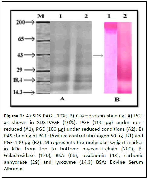
Current study evaluates the anticoagulant and antiplatelet activity of Pennisetum Glaucum Extract (PGE). PGE showed similar kind of protein banding pattern on 10% SDSPAGE, from the range 200 kDa to 14 kDa under reducing and non-reducing conditions. PGE was positive for PAS staining as it taken up the stain at the low molecular weight region (Figure 1). Furthermore, PGE hydrolysed casein with the specific activity of 0.2 Units/mg/min at 37°C, suggesting its proteolytic activity. The proteoltic activity of PGE was completely abolished by 1,10- Phenanthrolene but EGTA, EDTA, IAA and PMSF were insensitive, revealed the presence of zinc dependent metalloprotease (Table 1).
Figure 1: A) SDS-PAGE 10%; B) Glycoprotein staining. A) PGE as shown in SDS-PAGE (10%): PGE (100 μg) under nonreduced (A1), PGE (100 μg) under reduced conditions (A2). B) PAS staining of PGE: Positive control fibrinogen 50 μg (B1) and PGE 100 μg (B2). M represents the molecular weight marker in kDa from top to bottom: myosin-H-chain (200), β- Galactosidase (120), BSA (66), ovalbumin (43), carbonic anhydrase (29) and lysozyme (14.3) BSA: Bovine Serum Albumin.
| Inhibitor (5 mM each) | Activity/Residual activity |
|---|---|
| None | 90.25 |
| EDTA | 93.95 |
| EGTA | 83.2 |
| 1,10-Phenanthroline | 3.23 |
| IAA | 75.4 |
| PMSF | 84.09 |
Table 1: Effect of inhibitors on the proteolytic activity of PGE.
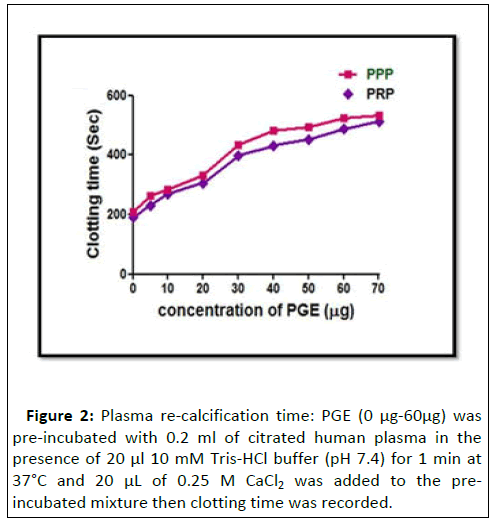
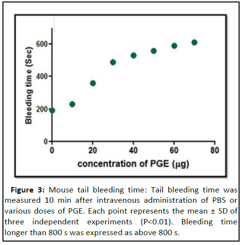
PGE increased the plasma coagulation time of both PRP and PPP from the control 210 s to 620 s and 246 s to 665 s respectively at the concentration of 60 μg, above the dose of 70 μg, it reached saturation (Figure 2). The exhibited anticoagulant property of PGE was also confirmed by in-vivo mouse tail bleeding assay (Figure 3). When PGE was injected intravenously, it significantly prolonged the mice tail bleeding time from PBS treated control 200 sec to 600 sec at the concentration of 70 μg. In addition, PGE prolonged only APTT but not PT revealed that its interference in intrinsic pathway of blood coagulation cascade (Table 2).
| PGE | PT clotting time in sec | PT (INR values) | APTT clotting in sec | APTT ratio |
|---|---|---|---|---|
| 0 | 11.2 ± 0.05 | 0.95 ± 0.02 | 40.3 ± 0.04 | 1.45 ± 0.01 |
| 4 | 11.3 ± 0.04 | 0.96 ± 0.08 | 59.9 ± 0.05 | 1.49 ± 10.03 |
| 8 | 11.5 ± 0.04 | 1.05 ± 0.02 | 75.9 ± 0.06 | 1.55 ± 0.02 |
| 12 | 11.7 ± 20.03 | 1.09 ± 10.01 | 89.7 ± 0.05 | 2.03 ± 0.07 |
Table 2: Effect of PGE on PT and APTT.
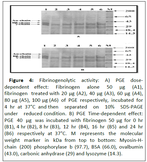
The proteolytic activity of PGE was also confirmed used fibrinogen and other plasma proteins. To the surprise, PGE cleaved only Aα and Bβ chain of human fibrinogen without affecting γ chain at the concentration of 100 μg at 37°C for 4hr incubation period. While, incubation time was increased up to 24 hr, PGE degraded partially γ chain of fibrinogen at the concentration of 40 μg (Figure 4).
Figure 4: Fibrinogenolytic activity: A) PGE dosedependent effect: Fibrinogen alone 50 μg (A1), fibrinogen treated with 20 μg (A2), 40 μg (A3), 60 μg (A4), 80 μg (A5), 100 μg (A6) of PGE respectively, incubated for 4 hr at 37°C and then separated on 10% SDS-PAGE under reduced condition. B) PGE Time-dependent effect: PGE 40 μg was incubated with fibrinogen 50 μg for 0 hr (B1), 4 hr (B2), 8 hr (B3), 12 hr (B4), 16 hr (B5) and 24 hr (B6) respectively at 37°C. M represents the molecular weight marker in kDa from top to bottom: Myosin-Hchain (200) phosphorylase b (97.7), BSA (66.0), ovalbumin (43.0), carbonic anhydrase (29) and lysozyme (14.3).
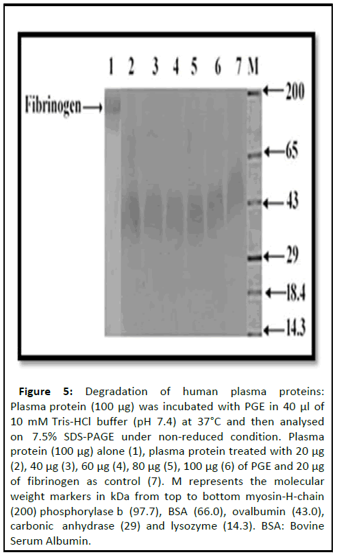
In order to understand the substrate speci icity of the PGE, it was incubated with plasma proteins. Interestingly, PGE did not degrade other plasma proteins but preferably degraded only ibrinogen, although for 12 hr incubation period at the concentration of 60 μg (Figure 5).
Figure 5: Degradation of human plasma proteins: Plasma protein (100 μg) was incubated with PGE in 40 μl of 10 mM Tris-HCl buffer (pH 7.4) at 37°C and then analysed on 7.5% SDS-PAGE under non-reduced condition. Plasma protein (100 μg) alone (1), plasma protein treated with 20 μg (2), 40 μg (3), 60 μg (4), 80 μg (5), 100 μg (6) of PGE and 20 μg of fibrinogen as control (7). M represents the molecular weight markers in kDa from top to bottom myosin-H-chain (200) phosphorylase b (97.7), BSA (66.0), ovalbumin (43.0), carbonic anhydrase (29) and lysozyme (14.3). BSA: Bovine Serum Albumin.
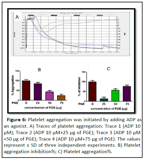
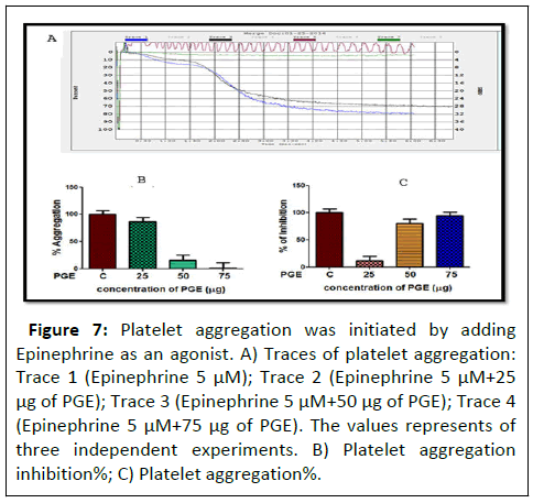
Moreover, PGE exhibited anti- platelet aggregation property by inhibiting ADP and epinephrine induced platelet aggregation (Figures 6 and 7). The obtained inhibition percentage was found to be 70% and 80% respectively at the concentration of 75 μg. Among the observed result PGE inhibited the agonists in the order epinephrine>ADP induced aggregation.
Figure 6: Platelet aggregation was initiated by adding ADP as an agonist. A) Traces of platelet aggregation: Trace 1 (ADP 10 μM); Trace 2 (ADP 10 μM+25 μg of PGE); Trace 3 (ADP 10 μM +50 μg of PGE); Trace 4 (ADP 10 μM+75 μg of PGE). The values represent ± SD of three independent experiments. B) Platelet aggregation inhibition%; C) Platelet aggregation%.
Figure 7: Platelet aggregation was initiated by adding Epinephrine as an agonist. A) Traces of platelet aggregation: Trace 1 (Epinephrine 5 μM); Trace 2 (Epinephrine 5 μM+25 μg of PGE); Trace 3 (Epinephrine 5 μM+50 μg of PGE); Trace 4 (Epinephrine 5 μM+75 μg of PGE). The values represents of three independent experiments. B) Platelet aggregation inhibition%; C) Platelet aggregation%.
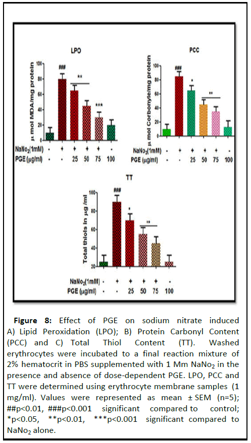
In addition, PGE was also analysed for its e icacy on oxidative stress. When the extent of lipid peroxidation was measured in terms of Malondialdehyde (MDA) levels as a marker of lipid peroxidation, there was a considerable increased level of MDA was observed in the sodium nitrate treated hemolysates. Interestingly, MDA level was normalized in PGE pre-incubated group. Whereas, the PGE treated group did not show any signi icant alterations (Figure 8).
Figure 8: Effect of PGE on sodium nitrate induced A) Lipid Peroxidation (LPO); B) Protein Carbonyl Content (PCC) and C) Total Thiol Content (TT). Washed erythrocytes were incubated to a final reaction mixture of 2% hematocrit in PBS supplemented with 1 Mm NaNo2 in the presence and absence of dose-dependent PGE. LPO, PCC and TT were determined using erythrocyte membrane samples (1 mg/ml). Values were represented as mean ± SEM (n=5); ##p<0.01, ###p<0.001 significant compared to control; *p<0.05, **p<0.01, ***p<0.001 significant compared to NaNo2 alone.
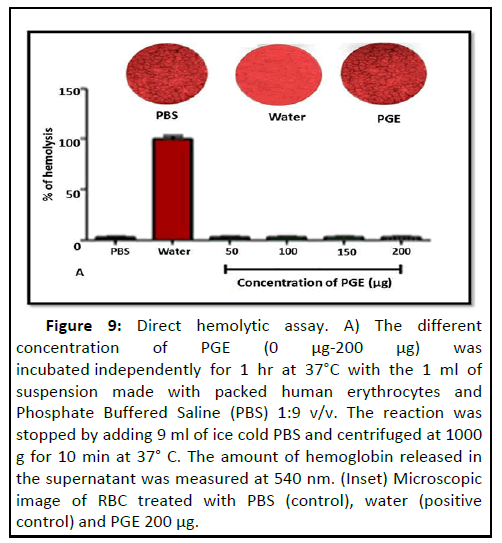
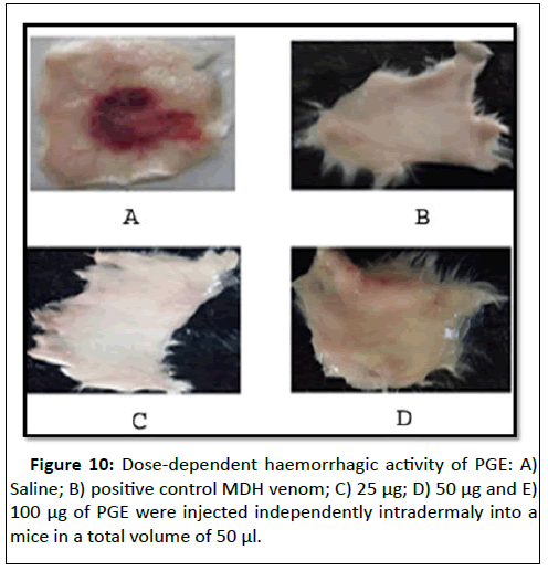
Furthermore, considerable elevation of Protein Carbonyl Content (PCC) in the sodium nitrate treated hemolysates was observed compared to the control group. However, in the preincubated group PGE re-established the altered PCC by reducing the levels towards normality. While, in the PGE-alone group the PCC levels remained unaltered. PGE significantly reduced the level of total thiol content compare to the sodium nitrate treated group. Fascinatingly, PGE did not lyse RBC; the microscopic image of RBC revealed that there was a massive destruction of RBC treated with water a positive control. While, PGE treated RBC cells were remained normal in their morphology, compared to the PBS treated RBC (Figure 9). PGE was devoid of haemorrhage and edema forming activities in experimental mice up to the concentration of 150 μg. Whereas, positive control Daboia russelli venom induced edema haemorrhage in experimental mice suggesting its non-toxic property (Figure 10).
Figure 9: Direct hemolytic assay. A) The different concentration of PGE (0 μg-200 μg) was incubated independently for 1 hr at 37°C with the 1 ml of suspension made with packed human erythrocytes and Phosphate Buffered Saline (PBS) 1:9 v/v. The reaction was stopped by adding 9 ml of ice cold PBS and centrifuged at 1000 g for 10 min at 37° C. The amount of hemoglobin released in the supernatant was measured at 540 nm. (Inset) Microscopic image of RBC treated with PBS (control), water (positive control) and PGE 200 μg.
Discussion
Eventhough, pearl millets (Pennisetum glaucum) are the store house of high quantity of nutritional and nutraceutical components, they are the most underutilized groups of cereals. Infact, they are yet considered as food of low income group. Researchers were documented the presence of proteins, unsaturated fatty acids, dietary fiber and secondary metabolites attributed in the prevention of many diseases such as, cardiovascular, diabetes and cataractogenesis. However, there are no reports on the beneficial role of proteins/proteolytic enzymes from the pearl millet. Hence, this study throws a light on the anticoagulant, antiplatelet and antioxidant properties of Pennisetum glaucum (pearl millet) protein extract. Oxidative stress is the one of the key contributors that causes hyper activation of coagulation factors and platelets results in thrombosis. Thrombosis is the key culprit in the pathophysiology of cardiovascular and cerebro vascular complications. Anticoagulant and antiplatelet agents have been extensively used in the treatment of thrombosis. While, fatal side effects such as, nausea, vomiting, headache, internal bleeding and birth defects limits their usage. Therefore, identifying the novel drug from natural sources with no side effects could help in the better controlling of thrombotic disorders. Thus, the current study deals with the anticoagulant and antiplatelet properties of PGE.
PGE showed similar protein banding pattern under reduced and non-reduced conditions revealed, it contains only monomeric proteins. Although, several protein bands were identified in the different molecular weight region, only one band was positive for PAS-staining. Hence, PGE has only one glycoprotein. PGE exhibited proteolytic activity and it was sensitive to 1,10-penanthrolene, suggesting the presence of zinc dependent metalloprotease. Ramana, et al., was detected the proteolytic activity in the germinated pearl millet. However, several researchers identified the protease inhibitors those exhibits antifungal and antibacterial effect from the pearl millet. Interestingly, PGE showed anticoagulant and antiplatelet properties. Hemostasis is a defence mechanism that involved in the arrest of bleeding at the site of injury. Three key events takes place during hemostasis namely vasoconstriction, platelet plug formation and clot formation. While, uncontrolled way of said mechanism leads to thrombosis. PGE showed anticoagulation in both PRP and PPP and mice tail bleeding time as well. In addition, PGE specifically prolonged only the activated partial thromboplastin time but not prothrombin time, suggests that observed anticoagulation of the PGE could be due to its interference in intrinsic pathway.
Blood coagulation cascade comprise of intrinsic, extrinsic pathway, where both the pathways converge at common site that involves the activation factor Xa, it converts prothrombin to thrombin in turn the later converts fibrinogen to fibrin. Site specific anticoagulants (factor Xa and thrombin inhibitors) with no side effects have better therapeutic value in the management of thrombosis. Furthermore, PGE non-specifically degraded all the chains of human fibrinogen supports its anticoagulation. Fibrinogenases those hydrolyzes Aα and Bβ chains of fibrinogen and generates fibrino peptide-A and B are thrombin like enzymes results in procoagulation activity. While, fibrinogenases hydrolyzes fibrinogen from C-terminal end generates condensed fibrinogen structure end up in anticoagulation activity. PGE did not hydrolyse other plasma proteins except fibrinogen suggests its substrate specificity.
PGE exhibited anti-platelet aggregation property by inhibiting ADP and epinephrine induced platelet aggregation. Platelet aggregation inhibition ability of PGE for both the agonists (ADP and epinephrine) seems to be similar. Antiplatelet effect was reported from jackfruit, bitter gourd and Pisum sativum seeds.
The redox state modulates the functions of several cells including platelet as well. Oxidative stress is associated with several diseases, namely, diabetes, cancer, cardiovascular neurodegenerative diseases and hemolytic anemias. Oxidative stress in platelets is the key contributing factor that leads to their hyper activation and excess clot formation, Hence, thromboembolic complications are common co-morbidity factors of said diseases. Lipid peroxidation and protein oxidation are considered to be the key events that indicate marked oxidative stress. PGE normalized the level of lipid peroxidation, Protein Carbonyl Content (PCC) and total thiol content in the experimental animals.
Conclusion
In conclusion, PGE possess zinc dependent proteolytic activity, exhibited anticoagulant, antiplatelet and non-toxic properties. Therefore, it could be a better contender to treat thrombotic disorders Suggesting its beneficial role in managing thrombotic complctaions. Fascinatingly, PGE exhibited non-toxic property as it was unable to cleave RBC and devoid of haemorrhage and edema forming activities in experimental animals.
Acknowledgment
D.S and M.S thank the department of science and technology, government of India, New Delhi and vision group on science and technology, government of Karnataka, Bangalore for financial assistance.
Conflict of Interest
The authors declared no potential conflict of interest with respect to the authorship and publication.
References
- Liu S, Manson JE, Lee IM, Cole SR, Hennekens CH, et al. (2000) Fruit and vegetable intake and risk of cardiovascular disease: The women's health study. Am J Clin Nutr 72: 922-928.
[Crossref] [Google Scholar] [PubMed]
- Joshipura KJ, Ascherio A, Manson JE, Stampfer MJ, Rimm EB, et al. (1999) Fruit and vegetable intake in relation to risk of ischemic stroke. Jama 282: 1233-1239.
[Crossref] [Google Scholar] [PubMed]
- Badau MH, Nkama I, Jideani IA (2005) Phytic acid content and hydrochloric acid extractability of minerals in pearl millet as affected by germination time and cultivar. Food Chem 92: 425-435.
- Blois MS (1958) Antioxidant determinations by the use of a stable free radical. Nature 181: 1199-1200.
- Velioglu Y, Mazza G, Gao L, Oomah BD (1998) Antioxidant activity and total phenolics in selected fruits, vegetables, and grain products. J Agric Food Chem 46: 4113-4117.
- Raj SN, Chaluvaraju G, Amruthesh KN, Shetty HS, Reddy MS, et al. (2003) Induction of growth promotion and resistance against downy mildew on pearl millet (Pennisetum glaucum) by rhizobacteria. Plant Dis 87: 380-384.
[Crossref] [Google Scholar] [PubMed]
- Bradford MM (1976) A rapid and sensitive method for the quantitation of microgram quantities of protein utilizing the principle of protein-dye binding. Anal Biochem 72: 248-254.
[Crossref] [Google Scholar] [PubMed]
- Leiman PG, Kanamaru S, Mesyanzhinov VV, Arisaka F, Rossmann MG, et al. (2003) Structure and morphogenesis of bacteriophage T4. Cell Mol Life Sci CMLS 60 2356-2370.
[Crossref] [Google Scholar] [PubMed]
- Leach BS, Collawn Jr JF, Fish WW (1980) Behavior of glycopolypeptides with empirical molecular weight estimation methods. 1. In sodium dodecyl sulfate. Biochemistry 19: 5734-5741.
[Crossref] [Google Scholar] [PubMed]
- Satake M, Murata Y, Suzuki T (1963) Studies on snake venom XIII. Chromatographic separation and properties of three proteinases from Agkistrodon halys blomhoffii venom. J Biochem 53: 438-447.
[Crossref] [Google Scholar] [PubMed]
- Pommelet, Durand, Laurian, Devictor (1998) Haemophilia A: Two cases showing unusual features at birth. Haemophilia 4: 122-125.
[Crossref] [Google Scholar] [PubMed]
- Ouyang C, Teng CM (1976) Fibrinogenolytic enzymes of Trimeresurus mucrosquamatus venom. Biochim Biophys Acta 420: 298-308.
[Crossref] [Google Scholar] [PubMed]
- Kumar MS, Devaraj VR, Vishwanath BS, Kemparaju K (2010) Anti-coagulant activity of a metalloprotease: further characterization from the Indian cobra (Naja naja) venom. J Thromb Thrombolysis 29: 340-348.
[Crossref] [Google Scholar] [PubMed]
Open Access Journals
- Aquaculture & Veterinary Science
- Chemistry & Chemical Sciences
- Clinical Sciences
- Engineering
- General Science
- Genetics & Molecular Biology
- Health Care & Nursing
- Immunology & Microbiology
- Materials Science
- Mathematics & Physics
- Medical Sciences
- Neurology & Psychiatry
- Oncology & Cancer Science
- Pharmaceutical Sciences
